18F-C2Am is a novel imaging agent that will identify apoptotic and necrotic cells.
18F-C2Am is radioactive, so is likely to be more sensitive than the 13C imaging agents and the hope is it would also be able to detect metastasis. 18F-C2Am detection is based on an increase in signal following treatment as more cells become apoptotic if the tumour is responding to treatment.
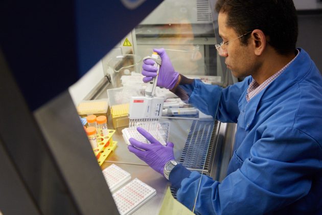
C2Am if proven successful, also has the potential to be used as a theragnostic. This could be imaged on PET/CT meaning that for patients in which MRI scans are contraindicated this could provide an alternative to fumarate. C2Am is expected to detect all forms of cell death and may provide a large window of opportunity for imaging following cell death. Due to the nature of the radioactive probe we hope that there is a good signal to noise ratio and the agent will confer sensitivity. C2Am, as a PET agent, can also be used for whole body imaging. It is currently being tested in multiple rodent models. The team, in collaboration with FUJIFILM Diosynth Biotechnologies is manufacturing a protein to GMP standards to be able to inject it into humans for the first-in-human testing.
18F-C2Am Infographic

Bulat, F., et al,18F-C2Am: a targeted imaging agent for detecting tumor cell death in vivo using positron emission tomography, EJNMMI Research, 9 Dec 2020.
Trialing novel cancer therapies in the clinic would benefit from imaging agents that can detect early evidence of treatment response. The timing, extent and distribution of cell death in tumors following treatment can give an indication of outcome. We describe here an 18F-labeled derivative of a phosphatidylserine-binding protein, the C2A domain of Synaptotagmin-I (C2Am), for imaging tumor cell death in vivo using PET.

The Mark Foundation Institute for Integrated Cancer Medicine (MFICM) at the University of Cambridge aims to revolutionise cancer care by affecting patients along their treatment pathway.


